
The Evolving Use of Intraoperative MRI in Brain Tumor Surgery
By Lily H. Kim MD, Michael Lim, MD, Professor and Chair of Neurosurgery at Stanford University School of Medicine, Stanford University School of Medicine
Brain tumor surgery poses a unique challenge, where the delicate balance between maximizing tumor removal while preventing deficits is paramount. In taking care of tumor patients, the goals are to improve survival while maintaining or improving quality of life. Conceptually, removing more tumor improves length of life; however, if a surgeon were to induce a neurological deficit, the benefit of improved survival is lost, in addition to compromising quality of life. Hence, for surgeons, there is a fine line at which one can be too aggressive in removing the tumor. To plan for a maximal safe resection, we as neurosurgeons carefully analyze preoperative imaging, which routinely employs state-of-the-art imaging, such as high-resolution brain magnetic resonance imaging (MRI) and vascular imaging. When the brain tumor involves areas of the brain especially important for neurological function (such as speech and movement), termed “eloquent” areas, advanced imaging techniques such as functional MRI and diffusion tensor imaging are also utilized to identify and create a detailed surgical roadmap and plan for resection.
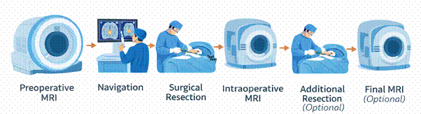
Imaging is important not only in the preoperative stage for planning but also during the operation for active guidance. In addition to allowing for planning, preoperative imaging can be used to provide a GPS for the brain using specially designed navigation systems. While this technology has allowed for smaller openings, the accuracy of the navigation system decreases during surgery due to the dynamic factors. In particular, variables such as loss of cerebrospinal fluid loss and decreased mass effect from tumor resection cause the brain to shift. In these cases, intraoperative magnetic resonance imaging (iMRI) offers an important adjunct to improve outcomes for our patients.
Given the continued need for precise and safe surgery, our field has always been open to incorporating innovative technologies to improve patient outcomes. And iMRI represents a major advancement in this continuous pursuit.
In addition to the need for continued accuracy of navigation, the true value of intraoperative imaging comes into play when assessment of residual tumor is visual. At points through the surgery, clear borders can be lost and as the cavity collapses, residual tumor may be lost from view. This is especially relevant in low-grade gliomas, in which surgical resection can be curative, but the subtle differences in tissue characteristics make it challenging to visually distinguish tumor margins from normal brain tissue. This contrasts with high-grade gliomas in which the tumor tissue is often distinctively different in appearance and texture, though iMRI can still be a useful adjunct to achieve maximal resection.
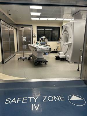
Although its development dates back to the 1990s, iMRI technology was not in widespread use due to various limitations, such as the physical constraints of operating around an MRI, sterility, and logistics associated with integrating large MRI scanners into operating suites. Furthermore, many surgical instruments (made of metal) and anesthetic equipment cannot be used near or in the magnet of the MRI. To overcome some of these hurdles, the MRI suites of today use two rooms. The MRI is put into another shielded room and then brought out on rails or in other configurations. The patient is moved into the room housing the MRI when imaging is needed. Currently, only a limited number of hospitals are equipped with a built-in intraoperative MRI suite. Another limitation is the high cost needed for both the initial setup and ongoing maintenance. Prolonged operative time is another drawback, with studies showing an addition of 45-90 minutes on average, which not only leads to increased costs but also potential patient risk due to extended anesthetic exposure. In addition, the need to sterilely re-drape the surgical site and higher personnel traffic could theoretically increase wound infection risk, though studies to date have not demonstrated such an increase. Moreover, operating room personnel must implement established MRI safety protocols, including verification that standard surgical and anesthetic equipment, such as the head fixation system, is MRI-compatible.
Despite these initial challenges, the adoption of iMRI technology has been linked to improved patient outcomes. Many studies have reported a higher likelihood of achieving gross total resection for both high-grade and low-grade gliomas when iMRI is utilized compared to conventional surgical techniques. Some studies have shown that when surgeons employed the iMRI, they ended up finding additional tumors that were left behind in as high as 60% of cases. This has been verified in randomized controlled trials, which is considered the highest level of evidence in medical research.
Future advancements in iMRI technology will include integrating artificial intelligence for immediate automated tumor margin and volumetric assessment to further aid surgical resection. If MRI sequences could be shortened and acquisition times minimized without compromising image quality, iMRI could be integrated more seamlessly into the current operative workflow. Additionally, the development of more compact and portable iMRI systems could expand their availability beyond academic centers and high-resource countries. Finally, augmented reality can further enhance this technology by superimposing real-time imaging directly onto the operative field and creating an immersive navigational experience for the surgeon.
Given the continued need for precise and safe surgery, our field has always been open to incorporating innovative technologies to improve patient outcomes. And iMRI represents a major advancement in this continuous pursuit. It is an exciting time for neurosurgery, as cutting-edge technology will only help us push the boundaries of possibilities in oncological treatment, offering hope for a better and longer life for our patients.
References
Ahmed, A., Shakir, M., Malik, M. J. A., Arif, A., Islam, O., Mubarak, F., Knopp, E., Siddiqui, K., & Enam, S. A. (2023). Intraoperative use of low-field magnetic resonance imaging for brain tumors: A systematic review [Review of Intraoperative use of low-field magnetic resonance imaging for brain tumors: A systematic review]. Surgical Neurology International, 14, 357. Medknow. https://doi.org/10.25259/sni_510_2023
Akbari, S. H. A., Sylvester, P., Kulwin, C., Shah, M. V., Somasundaram, A., Kamath, A. A., Beaumont, T., Rich, K. M., & Chicoine, M. R. (2018). Initial Experience Using Intraoperative Magnetic Resonance Imaging During a Trans-Sulcal Tubular Retractor Approach for the Resection of Deep-Seated Brain Tumors: A Case Series. Operative Neurosurgery, 16(3), 292. https://doi.org/10.1093/ons/opy108
Bunyaratavej, K., Siwanuwatn, R., Tuchinda, L., & Wangsawatwong, P. (2022). Impact of Intraoperative Magnetic Resonance Imaging (i-MRI) on Surgeon Decision Making and Clinical Outcomes in Cranial Tumor Surgery. Asian Journal of Neurosurgery, 17(2), 218. https://doi.org/10.1055/s-0042-1751008
Fujii, Y., Ogiwara, T., Watanabe, G., Hanaoka, Y., Goto, T., Hongo, K., & Horiuchi, T. (2021). Intraoperative Low-Field Magnetic Resonance Imaging-Guided Tumor Resection in Glioma Surgery: Pros and Cons. Journal of Nippon Medical School, 89(3), 269. https://doi.org/10.1272/jnms.jnms.2022_89-301
Gárzón-Muvdi, T., Kut, C., Li, X., & Chaichana, K. L. (2017). Intraoperative imaging techniques for glioma surgery [Review of Intraoperative imaging techniques for glioma surgery]. Future Oncology, 13(19), 1731. Future Medicine. https://doi.org/10.2217/fon-2017-0092
Hollon, T., Hervey‐Jumper, S. L., Sagher, O., & Orringer, D. A. (2015). Advances in the Surgical Management of Low-Grade Glioma [Review of Advances in the Surgical Management of Low-Grade Glioma]. Seminars in Radiation Oncology, 25(3), 181. Elsevier BV. https://doi.org/10.1016/j.semradonc.2015.02.007
Hu, S., Kang, H., Baek, Y., Fakhri, G. E., Kuang, A., & Choi, H. S. (2018). Real‐Time Imaging of Brain Tumor for Image‐Guided Surgery [Review of Real‐Time Imaging of Brain Tumor for Image‐Guided Surgery]. Advanced Healthcare Materials, 7(16). Wiley. https://doi.org/10.1002/adhm.201800066
Jólesz, F. A. (2010). Intraoperative Imaging in Neurosurgery: Where Will the Future Take Us? Acta Neurochirurgica. Supplementum, 21. https://doi.org/10.1007/978-3-211-99651-5_4
Lam, F. C., Tsedev, U., Kasper, E. M., & Belcher, A. M. (2022). Forging the Frontiers of Image-Guided Neurosurgery—The Emerging Uses of Theranostics in Neurosurgical Oncology. Frontiers in Bioengineering and Biotechnology, 10. https://doi.org/10.3389/fbioe.2022.857093
Lv, X., Wang, G., Wang, J. Z., & Wu, Z. (2021). Frontiers in Clinical Neurosurgery. In IntechOpen eBooks. IntechOpen. https://doi.org/10.5772/intechopen.92911
Navarro-Olvera, J. L., Parra-Romero, G., Heres-Becerril, S. D., Trejo‐Martínez, D., Ruiz, J. D. C., Echeverría, K. G., & Aguado-Carrillo, G. (2021). Management of Brain Tumors in Eloquent Areas with Awake Patient. In IntechOpen eBooks. IntechOpen. https://doi.org/10.5772/intechopen.95584
Olubiyi, O. I., Özdemir, A., Incekara, F., Tie, Y., Dolati, P., Hsu, L., Santagata, S., Chen, Z., Rigolo, L., & Golby, A. J. (2015). Intraoperative Magnetic Resonance Imaging in Intracranial Glioma Resection: A Single-Center, Retrospective Blinded Volumetric Study. World Neurosurgery, 84(2), 528. https://doi.org/10.1016/j.wneu.2015.04.044 Senft, C., Bink, A., Franz, K., Vatter, H., Gasser, T., & Seifert, V. (2011). Intraoperative MRI guidance and extent of resection in glioma surgery: a randomised, controlled trial. The Lancet Oncology, 12(11), 997. https://doi.org/10.1016/s1470-2045(11)70196-6
Sharma, M., Wang, D., Palmisciano, P., Ugiliweneza, B., Woo, S. Y., Nelson, M., Miller, D. M., Savage, J. J., Boakye, M., Andaluz, N., Mistry, A. M., Chen, C. C., & Williams, B. J. (2022). Is intraoperative MRI use in malignant brain tumor surgery a health care burden? A matched analysis of MarketScan Database. Journal of Neuro-Oncology, 160(2), 331. https://doi.org/10.1007/s11060-022-04142-0
Zhang, W., Ille, S., Schwendner, M., Wiestler, B., Meyer, B., & Krieg, S. M. (2023). The Impact of ioMRI on Glioblastoma Resection and Clinical Outcomes in a State-of-the-Art Neuro-Oncological Setup. Cancers, 15(14), 3563. https://doi.org/10.3390/cancers15143563