The Promise of Spatial Analysis for Medical Research
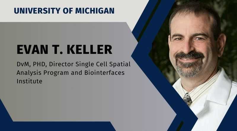
By Evan T. Keller DVM, PhD, Director Single Cell Spatial Analysis Program and Biointerfaces Institute, University of Michigan, Ann Arbor, Michigan
When a patient presents with a challenging disorder or a tumor, it is often important to evaluate the diseased tissue to help define that patient’s illness. This has been accomplished for over a hundred years using histological evaluation, i.e., by looking at the tissue under the microscope. Specifically, the tissue is processed so that a thin slice can be evaluated using a microscope to see the cellular structure, and the characteristics of the cells and the tissue structure are examined by a trained pathologist. These characteristics, such as cell shape and size, provide clues as to the processes occurring in the tissue. To provide further information, the tissues are often stained with different dyes to help identify different cells, their structures and specific proteins. While these methods have served well for many years, they only allow evaluation of a limited number of parameters and provide limited information. This is of particular importance in current times due to the identification of many subtypes of disease and subtypes of cancer. Recently, breakthroughs in molecular methods to evaluate the expression from 100s to 1000s of genes in tissues, termed spatial transcriptomics, have provided novel methods to glean large amounts of information from tissues. This allows for the identification of subtypes, which can lead to diagnosis and prognosis and potentially predict drug response, allowing for enhancing precision medicine.
As spatial analytic methods develop, novel ways to analyze them will be applied to enhance our understanding of the data they generate. Artificial Intelligence (AI) will greatly facilitate the utility and application of spatial transcriptomics.
Spatial transcriptomics is an exciting new technique in the field of molecular biology that allows researchers to study gene activity in various regions of tissues. Unlike traditional methods that analyze gene expression in bulk samples, spatial transcriptomics retains spatial context, meaning it allows scientists to see which genes are active in specific cells or regions of a tissue sample. This is crucial for understanding complex biological processes and diseases, as it helps pinpoint where and how genes are functioning differently in, for example, diseased versus healthy tissue. By capturing the spatial organization of gene expression, researchers can gain insights into the cellular architecture and interactions within tissues, which is particularly valuable for studying diseases like cancer, where the tumor microenvironment plays a critical role (Ståhl et al., 2016; Asp et al., 2021).
Several different methodologies have been used to evaluate spatial gene expression. These can be broadly divided into either sequencing-based or imaging-based. In terms of sequencing-based, tissue samples are first sectioned and placed on specially designed slides that have spatially barcoded spots. Each spot contains capture oligos with unique molecular barcodes that capture RNA molecules from the tissue section. The tissue is then stained to visualize its structure and subsequently treated to release RNA, which binds to the barcodes. In the next step, the captured RNA is sequenced to identify the gene activity at each spot on the slide. These data are then combined with imaging data to create a comprehensive map showing where each gene is active within the tissue. A limitation of this method is that it can be challenging to clearly align the transcripts with cells due to the nature of the capture oligo alignment.
In contrast to the sequencing-based methods, image-based methods involve a process in which a library of probes is cyclically incubated (a few probes at a time) on a tissue and imaged (typically fluorescently). A strength of this method is that imaging allows for true identification of the cells, and, subsequently, clearly aligns the RNA transcripts in the cells. These techniques are powerful not only for their ability to provide a high-resolution view of gene expression but also for their potential to revolutionize our understanding of tissue organization and pathology (Ståhl et al., 2016; Maniatis et al., 2019; Burgess, 2019; Chen et al., 2020; Marx, 2021).
As spatial analytic methods develop, novel ways to analyze them will be applied to enhance our understanding of the data they generate. Artificial Intelligence (AI) will greatly facilitate the utility and application of spatial transcriptomics. By leveraging AI algorithms, especially machine learning and deep learning, scientists can process vast amounts of complex data more efficiently than ever before. These algorithms can identify patterns, correlations, and unique gene expression signatures within the spatial context of tissues that will take advantage of clinical correlatives. This enables researchers to gain deeper insights into cellular functions, developmental processes, and disease mechanisms. For instance, AI can help pinpoint how certain genes are expressed differently in healthy versus diseased tissues and uncover new therapeutic targets. In essence, in the context of spatial analysis, AI has great potential to acta as a powerful tool to translate intricate spatial transcriptomic data into actionable clinical understanding.
References:
- Ståhl, P. L., Salmén, F., Vickovic, S., Lundmark, A., Navarro, J. F., Magnusson, J., … & Frisen, J. (2016). Visualization and analysis of gene expression in tissue sections by spatial transcriptomics. Science, 353(6294), 78-82.
- Asp, M., Bergenstråhle, J., Lundeberg, J. (2021). Spatially resolved transcriptomes—next generation tools for tissue exploration. BioEssays, 43(5), e2000221.
- Maniatis, S., Aijo, T., Vickovic, S., Braine, C., Kang, K., Mollbrink, A., … & Lundeberg, J. (2019). Spatiotemporal dynamics of molecular pathology in amyotrophic lateral sclerosis. Science, 364(6435), 89-93.
- Burgess, D. J. (2019). Spatial transcriptomics coming of age. Nature Reviews Genetics, 20(6), 307.
- Chen A, Liao S, Cheng M, Ma K, Wu L, Lai Y, Qiu X, Yang J, Xu J, Hao S, Wang X, Lu H, Chen X, Liu X, Huang X, Li Z, Hong Y, Jiang Y, Peng J, Liu S, Shen M, Liu C, Li Q, Yuan Y, Wei X, Zheng H, Feng W, Wang Z, Liu Y, Wang Z, Yang Y, Xiang H, Han L, Qin B, Guo P, Lai G, Muñoz-Cánoves P, Maxwell PH, Thiery JP, Wu QF, Zhao F, Chen B, Li M, Dai X, Wang S, Kuang H, Hui J, Wang L, Fei JF, Wang O, Wei X, Lu H, Wang B, Liu S, Gu Y, Ni M, Zhang W, Mu F, Yin Y, Yang H, Lisby M, Cornall RJ, Mulder J, Uhlén M, Esteban MA, Li Y, Liu L, Xu X, Wang J. Spatiotemporal transcriptomic atlas of mouse organogenesis using DNA nanoball-patterned arrays. Cell. 2022 May 12;185(10):1777-1792.e21. doi: 10.1016/j.cell.2022.04.003. Epub 2022 May 4. PMID: 35512705.
- Marx, V. (2021). Method of the Year: spatially resolved transcriptomics. Nature Methods, 18(1), 9-14.

