
The Role of Immersive Technologies in Clinical Anatomy Education
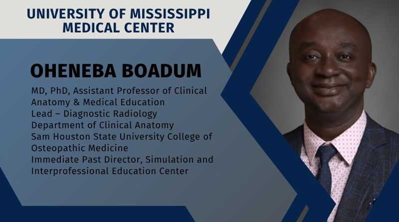
By Oheneba Boadum, MD, PhD, Assistant Professor of Clinical Anatomy & Medical Education, Lead – Diagnostic Radiology, Department of Clinical Anatomy, Sam Houston State University College of Osteopathic Medicine | Immediate Past Director, Simulation and Interprofessional Education Center, University of Mississippi Medical Center
Clinical anatomy has traditionally been taught through cadaveric dissection, a method that remains foundational in medical education [1]. Beyond anatomical knowledge, the dissection laboratory introduces students to professionalism, teamwork, and ethical responsibility [2]. Anatomical donors are often described as students’ first patients, shaping attitudes toward respect, empathy, and clinical accountability [3]. While cadaver-based anatomy can be emotionally challenging, it remains a formative experience that prepares learners for clinical practice [4].
Over the past decade, anatomy education has evolved with the integration of immersive technologies such as virtual reality (VR), augmented reality (AR), mixed reality (MR), and large- format digital dissection tables collectively referred to as extended reality (XR) [5]. Depending on curricular philosophy and institutional resources, medical schools now employ a range of instructional models, including cadaveric dissection, prosection, plastinated specimens, digital platforms, or hybrid approaches supplemented by XR.
Based on my experience as a clinical anatomist and medical educator, VR, AR, and MR function best as supplementary tools rather than replacements for traditional cadaveric learning. One of their most practical advantages is access. Anatomy laboratories are not always available outside scheduled hours, and prolonged time with donors is neither feasible nor desirable. XR allows students to revisit anatomical regions, consolidate learning, and clarify spatial relationships from home or other non-laboratory settings.
Used wisely, immersive technologies can extend the reach of anatomy education while preserving its human foundations.
Current Applications and Trends in Anatomy Education
In my teaching practice, XR has been most effective in elective settings and advanced learner contexts. Fourth-year medical students, residents, and practicing clinicians seeking anatomical refreshers often prefer VR, AR, or MR platforms over returning to the dissection laboratory [6]. These tools provide flexibility while maintaining anatomical rigor.
Certain anatomical regions consistently benefit from XR-based exploration. Students frequently rely on VR and MR visualization for the head and neck, pelvic anatomy, and complex neurovascular territories, areas traditionally recognized as challenging during cadaveric dissection [7]. These platforms allow learners to isolate structures, manipulate depth, and visualize spatial relationships that may be difficult to appreciate in donors due to preservation limitations or anatomical variation [8]. For some learners, immersive visualization supports spatial reasoning before or after dissection.
Beyond gross anatomy, XR plays an important role in procedural education. Simulation using task trainers and digital environments is now central to teaching skills such as lumbar puncture, thoracentesis, and central venous access [4]. VR-based procedural simulations allow repeated practice in low-risk environments, helping interns and junior trainees develop confidence and coordination prior to patient encounters [4].
I have also been involved in projects using photogrammetry to create detailed three-dimensional anatomical models. These reconstructions allow learners to zoom, rotate, and interrogate anatomy in ways not always possible with physical specimens, reinforcing spatial understanding and revealing perspectives that may be difficult to access in the laboratory [9].
Educational Value Beyond the Laboratory
Immersive technologies offer strong potential for pre-laboratory preparation and post-laboratory consolidation. Structured pre- and post-lab guidance improves learning when students arrive oriented and leave with opportunities for efficient review [5]. Mixed reality sessions before cadaver lab can reduce cognitive load during dissection, while post-lab VR review supports spaced repetition and reinforcement of key concepts [6].
Importantly, XR contributes to equity and access. Institutions with limited cadaver availability, constrained laboratory space, or geographic barriers can use immersive tools to broaden anatomical exposure [10]. Learners across regions may gain standardized access to complex spatial tasks and rare anatomical variations, helping to democratize anatomy education without eliminating the ethical and professional foundations of donor-based learning [11].
Challenges in Adoption and Implementation
Despite their promise, XR technologies present notable challenges. Cost and sustainability remain major barriers. Head-mounted displays, enterprise software licenses, and high-quality 3D content require substantial investment and ongoing technical support. These tools also demand dedicated physical space, audiovisual infrastructure, and institutional governance; resources often underestimated during planning.
Faculty development is another critical hurdle. XR technologies are pedagogical tools, not educational outcomes. Without clear curricular alignment and assessment mapping, they risk becoming superficial add-ons. Successful integration requires structured lesson plans explicitly linking immersive interaction to learning objectives and assessment blueprints. Both faculty and learners face learning curves, which can be difficult to accommodate within compressed anatomy curricula.
Content fidelity and standardization pose additional concerns. Not all commercially available 3D models adhere strictly to anatomical nomenclature or accurately represent anatomical variation. Educators must critically curate content rather than assume equivalence across platforms.
Physiological and logistical issues must also be addressed. Some learners experience dizziness or motion discomfort in immersive environments. Headset hygiene, device sharing, and learner safety, particularly in fully immersive VR, require clear protocols and supervision. These elements support the requirement for specific areas and organized execution.
Positive and Negative Impacts of Emerging Technology
When thoughtfully implemented, XR offers clear benefits. Learner engagement is often high, and immersive rehearsal can be efficient, particularly for focused spatial or procedural tasks [4].Adjustable visualization, layered labeling, and interactive control can enhance accessibility for learners with diverse needs [6]. Additionally, XR platforms can standardize learning experiences when cadaver quality or access varies [10].
However, negative impacts must be acknowledged. Poorly aligned implementation can lead to cognitive overload, superficial engagement, or distraction from core learning goals. Hidden costs include faculty time, technical maintenance, and content curation persist long after initial acquisition. Evidence supporting XR remains variable across contexts, emphasizing the need for local evaluation and scholarly dissemination of outcomes [4].
Looking Forward
Immersive technologies are not redefining anatomy education by replacing cadaveric learning. Rather, they are reshaping how anatomical knowledge is accessed, reinforced, and applied. Their strengths lie in visualization, flexibility, and adaptability. The future of clinical anatomy Education will be intentionally hybrid, guided by pedagogy, ethics, and learner needs.
Used wisely, immersive technologies can extend the reach of anatomy education while preserving its human foundations. The challenge for educators and institutions is not whether to adopt these technologies, but how to integrate them responsibly, sustainably, and in service of meaningful learning outcomes. As evidence matures and costs decline, XR will likely become a standard adjunct in anatomy education worldwide.
References
- Stojanovska, M., Tingle, G., Tan, L., Ulrey, L., Simonson-Shick, S., Mlakar, J., Eastman, H., Gotschall, R., Boscia, A., Enterline, R., Henninger, E., Herrmann, K. A., Simpson, S. W., Griswold, M. A., & Wish-Baratz, S. (2019). Mixed Reality Anatomy Using Microsoft HoloLens and Cadaveric Dissection: A Comparative Effectiveness Study. Medical science educator, 30(1), 173–178. https://doi.org/10.1007/s40670-019-00834-x
- Wish-Baratz, S., Crofton, A. R., Gutierrez, J., Henninger, E., & Griswold, M. A. (2020). Assessment of Mixed-Reality Technology Use in Remote Online Anatomy Education. JAMA network open, 3(9), e2016271. https://doi.org/10.1001/jamanetworkopen.2020.16271
- Veer, V., Phelps, C., & Moro, C. (2022). Incorporating Mixed Reality for Knowledge Retention in Physiology, Anatomy, Pathology, and Pharmacology Interdisciplinary Education: A Randomized Controlled Trial. Medical science educator, 32(6), 1579–1586. https://doi.org/10.1007/s40670-022-01635-5
- Mao, R. Q., Lan, L., Kay, J., Lohre, R., Ayeni, O. R., Goel, D. P., & Sa, D. (2021). Immersive Virtual Reality for Surgical Training: A Systematic Review. The Journal of surgical research, 268, 40–58. https://doi.org/10.1016/j.jss.2021.06.045
- Zingg, L., Häusler, M., Hein, J., Jecklin, S., Kottner, S., Gascho, D., Cavalcanti, N., Voinov, P., Götschi, T., Carrillo, F., Lagler, F., Fürnstahl, P., & Farshad, M. (2025). Using Hologram-Based Augmented Reality in Anatomy Learning: The TEACHANATOMY Randomized Trial. Academic medicine : journal of the Association of American Medical Colleges, 100(6), 695–704. https://doi.org/10.1097/ACM.0000000000006012
- Lake K, McKittrick A, Desselle M, Padilha Lanari Bo A, Abayasiri RAM, Fleming J, et al. Cybersecurity and privacy issues in extended reality health care applications: Scoping review. JMIR XR Spatial Comput. 2024;1:e59409. URL: https://xr.jmir.org/2024/1/e59409; doi: 10.2196/59409
- U.S. Food and Drug Administration. Digital Health Center of Excellence [Internet]. 2024 [cited 2025 Dec 17]. Available from: https://www.fda.gov/medical-devices/digital-health- center-excellence
- Moro, C., Birt, J., Stromberga, Z., Phelps, C., Clark, J., Glasziou, P., & Scott, A. M. (2021). Virtual and Augmented Reality Enhancements to Medical and Science Student Physiology and Anatomy Test Performance: A Systematic Review and Meta-Analysis. Anatomical sciences education, 14(3), 368–376. https://doi.org/10.1002/ase.2049
- Tullos N, Eldridge J, Schaefer A, Norcross E, Boadum O, Thompson K, et al. Renal anatomy review activity using 3D digital models and clinical cases. Anat Sci Educ. 2025;00:1–9. https://doi.org/10.1002/ase.70151
- Tene T, Vique López DF, Valverde Aguirre PE, Orna Puente LM, Vacacela Gomez C. Virtual reality and augmented reality in medical education: an umbrella review. Front Digit Health. 2024 Mar 14;6:1365345. doi: 10.3389/fdgth.2024.1365345. PMID: 38550715; PMCID: PMC10973128.
- WHO. WHO guidelines on digital health interventions [Internet]. 2023 [cited 2025 Dec 17]. Available from: https://www.who.int/publications/i/item/9789241550505