
AI in Epilepsy Care: From Seizure Detection to Safer Clinical Decisions
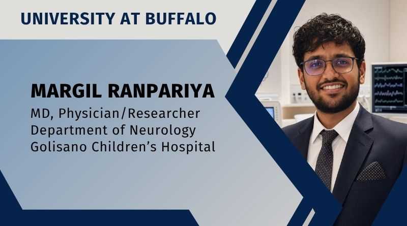
By Margil Ranpariya, MD, Physician/Researcher, Department of Neurology, University at Buffalo | Golisano Children’s Hospital
Epilepsy care is built around uncertainty. A patient may have a seizure at night and not remember it. A caregiver may miss a brief event. A routine electroencephalogram (EEG) may be normal even when the clinical story strongly suggests epilepsy. A surgeon may know that resection offers a real chance of seizure freedom, while also knowing that outcomes are not guaranteed. These are not small problems. They shape diagnosis, treatment, safety, and the daily confidence of people living with epilepsy.
Artificial intelligence (AI) is beginning to enter this difficult space, not as a replacement for neurologists, but as a second layer of pattern recognition. It is useful for analyzing lengthy signals, merging intricate inputs, and highlighting details that could otherwise go unnoticed. The most useful future for AI in epilepsy will not be a machine making isolated decisions. It will be a clinical system where human judgment remains central and AI helps make that judgment better informed.[1,2]
One of the clearest areas is EEG interpretation. EEG is one of the most important tools in epilepsy diagnosis, yet its interpretation requires expertise that is unevenly available. Even in advanced health systems, EEG workload is rising and inter-reader variability remains a concern. The SCORE-AI study showed that a convolutional neural network trained on 30,493 routine EEGs and tested on independent datasets could classify routine EEG abnormalities with performance similar to human experts, with reported area under the receiver operating characteristic curve values between 0.89 and 0.96.[3] That does not mean the EEG physician becomes unnecessary. It means that a validated system may help triage normal studies, support readers in under-resourced settings, and reduce avoidable misinterpretation.[3]
The goal is not to let algorithms speak for patients, but to help clinicians hear the hidden patterns in their disease.
Wearable seizure detection is another practical step. Many patients and families want objective seizure counts because self-reporting is imperfect. Studies cited in the International League Against Epilepsy–International Federation of Clinical Neurophysiology guideline show that 47%–63% of seizures in video-EEG monitoring units may be unrecognized by patients, with even higher rates for nocturnal seizures.[4] Wearable devices are most supported for generalized tonic-clonic and focal-to-bilateral tonic-clonic seizures. The guideline recommends clinically validated devices for selected patients, especially where an alarm can lead to rapid intervention. At the same time, it does not recommend current devices for routine clinical use across all other seizure types.[4] This distinction is important. A good epilepsy technology must be honest about what it can and cannot detect.
The next step is seizure forecasting. Detection says, “A seizure may be happening now.” Forecasting tries to say, “Your risk may be higher during this period.” That could change daily life. A patient might avoid swimming alone, driving, climbing, cooking over heat, or missing medication during a high-risk window. But forecasting should be treated like a weather forecast, not a promise. Wearables, diaries, sleep data, heart rate, electrodermal activity, stress, mood, and long-term seizure cycles may all contribute.[5] The evidence is promising, but long-term studies with EEG confirmation are still needed.[5] False alarms, privacy concerns, battery life, comfort, and user acceptance will decide whether patients keep using these tools after the first few weeks.[5]
AI may also improve care for people with drug-resistant epilepsy. Surgery can be life-changing for selected patients, but predicting seizure outcome remains difficult. A 2024 study of 294 patients undergoing temporal lobe resection reported that machine learning using five minutes of peri-ictal scalp EEG achieved high predictive performance for postoperative seizure control.[6] The authors also reported that an EEG-augmented model could reduce unsuccessful resections compared with a clinical-variable model.[6] This is a meaningful direction because scalp EEG is already part of the routine presurgical evaluation. However, such models need external validation, prospective testing, and careful integration into surgical conferences before they can influence major treatment decisions.
The same logic applies to medication choice, comorbidity prediction, imaging review, and remote monitoring. AI may eventually help identify medication response patterns, flag patients who need earlier referral to an epilepsy center, detect subtle magnetic resonance imaging lesions, or combine EEG and imaging into a more complete map of the epileptic network.[1,2] The future clinic may not depend only on a short visit every few months. It may include continuous but selective data streams, summarized in a way that is useful to both patient and clinician.[1,2,5]
There are risks. AI systems can carry bias if they are trained on narrow populations. They can fail when used with different EEG machines, different age groups, different seizure types, or low-quality data. They can create false confidence if a clinician treats a probability as a fact. They can widen disparities if only wealthy centers can afford the tools. They can also raise difficult questions about ownership of EEG, imaging, wearable, and electronic health record data.[1,2,5] For epilepsy, where stigma and privacy already matter deeply, these concerns are not technical footnotes. They are central to trust.
The most responsible future is therefore “decision intelligence”: building tools around real clinical questions, patient priorities, local resources, and measurable outcomes.[1] That means involving people with epilepsy, caregivers, neurologists, EEG technologists, engineers, ethicists, and health systems early. It also means testing models outside the hospital where they were built, monitoring performance after deployment, and being willing to withdraw tools that do not improve care.[1,2]
AI in epilepsy should be judged by practical standards: Does it shorten the time to diagnosis? Does it reduce missed seizures? Does it help identify surgical candidates earlier? Does it improve safety without overwhelming families with alarms? Does it work in low-resource settings, not only academic centers? Does it preserve dignity, privacy, and clinician accountability?[1,2,4]
The future of epilepsy care will still depend on listening carefully to patients. AI will not replace that conversation. Its best role is to bring better evidence into the room.
“The goal is not to let algorithms speak for patients, but to help clinicians hear the hidden patterns in their disease.”
References
- Josephson CB, Beniczky S, Denaxas S, et al. A call for ethical, equitable, and effective artificial intelligence to improve care for all people with epilepsy: a roadmap. A report by the ILAE Global Advocacy Council and Big Data Commission. Epilepsia. 2026;67:1555–1572. doi:10.1002/epi.70058
- Wang C, Yuan X, Jing W. Artificial intelligence in electroencephalography analysis for epilepsy diagnosis and management. Front Neurol. 2025;16:1615120. doi:10.3389/fneur.2025.1615120
- Tveit J, Aurlien H, Plis S, et al. Automated interpretation of clinical electroencephalograms using artificial intelligence. JAMA Neurol. 2023;80(8):805–812. doi:10.1001/jamaneurol.2023.1645
- Beniczky S, Wiebe S, Jeppesen J, et al. Automated seizure detection using wearable devices: a clinical practice guideline of the International League Against Epilepsy and the International Federation of Clinical Neurophysiology. Epilepsia. 2021;62:632–646. doi:10.1111/epi.16818
- Brinkmann BH, Karoly PJ, Nurse ES, et al. Seizure diaries and forecasting with wearables: epilepsy monitoring outside the clinic. Front Neurol. 2021;12:690404. doi:10.3389/fneur.2021.690404
- Sheikh SR, McKee ZA, Ghosn S, et al. Machine learning algorithm for predicting seizure control after temporal lobe resection using peri-ictal electroencephalography. Sci Rep. 2024;14:21771. doi:10.1038/s41598-024-72249-7